884:
505:
440:
363:
456:
532:
471:
56:
520:
40:
883:
355:
367:
than 2.5 mm. In the middle photo, the pupil is midway between the sclera posteriorly and the cornea anteriorly, indicating an EZ ratio of 0.5, and a medium chamber depth of about 2.5 mm. In the rightmost photo, the pupil is very anterior (forward), indicating an EZ ratio of more than 0.5 and a shallow anterior chamber of less than 2.5 mm.
366:
Figure 2. Different anterior chamber depths as seen from the lateral perpendicular (profile) view. The more forward the pupil is, the shallower the anterior chamber. In the leftmost photo, the pupil is relatively posterior (set back), indicating an EZ ratio of < 0.5 and an anterior chamber deeper
273:
The EZ ratio method is one way to calculate the estimated anterior chamber depth. To start, the patient looks at a target in the distance with one eye covered. The examiner takes a digital photograph of the open, examined eye, from the side, perpendicular to the visual axis (a profile photograph).
919:
1:posterior segment 2:ora serrata 3:ciliary muscle 4:ciliary zonules 5:Schlemm's canal 6:pupil 7:anterior chamber 8:cornea 9:iris 10:lens cortex 11:lens nucleus 12:ciliary process 13:conjunctiva 14:inferior oblique muscule 15:inferior rectus muscule 16:medial rectus muscle 17:retinal arteries and
470:
201:
are three main pathologies in this area. In hyphema, blood fills the anterior chamber as a result of a hemorrhage, most commonly after a blunt eye injury. Anterior uveitis is an inflammatory process affecting the
920:
veins 18:optic disc 19:dura mater 20:central retinal artery 21:central retinal vein 22:optic nerve 23:vorticose vein 24:bulbar sheath 25:macula 26:fovea 27:sclera 28:choroid 29:superior rectus muscle 30:retina
262:
250:
Determining the anterior chamber depth (ACD) is important in estimating the risk of angle closure glaucoma. There are various methods of measuring ACD, including examination through a
439:
707:
233:
The depth of the anterior chamber of the eye varies between 1.5 and 4.0 mm, averaging 3.0 mm. It tends to become shallower at older age and in eyes with
903:
531:
137:
900:
1163:
901:
906:
504:
915:
902:
907:
455:
887:
113:
261:
A simpler clinical method of quantitatively estimating ACD using smartphone photography (EZ ratio) was developed by Dr Ehud Zamir from the
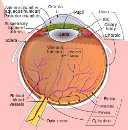
893:
711:
898:
362:
891:
910:
909:
763:
587:
890:
889:
1409:
897:
519:
908:
899:
896:
610:"A novel method of quantitative anterior chamber depth estimation using temporal perpendicular digital photography"
17:
905:
904:
888:
735:
911:
558:
376:
One peculiar feature of the anterior chamber is dampened immune response to allogenic grafts. This is called
132:
912:
1354:
1322:
1159:
1276:
1268:
948:
548:
237:(far sightedness). As depth decreases below 2.5 mm, the risk for angle closure glaucoma increases.
1460:
1103:
1093:
914:
895:
166:
756:
913:
916:
785:
96:
894:
1098:
1054:
659:"Induction of anterior chamber-associated immune deviation requires an intact, functional spleen"
1238:
1131:
862:
553:
144:
120:
108:
892:
1414:
1219:
1143:
1081:
1069:
1024:
348:
255:
1404:
749:
420:
219:
347:
of +/– 0.33 mm error, when compared to measurements of the anterior chamber depth by
8:
1429:
1135:
1086:
1074:
1064:
1019:
953:
867:
814:
484:
425:
344:
215:
186:
741:
55:
1399:
1179:
1175:
1171:
1119:
958:
842:
809:
684:
658:
634:
609:
480:
381:
330:
258:
photography. These methods require sophisticated examination equipment and expertise.
1439:
1155:
1059:
976:
847:
773:
689:
639:
583:
278:
738:
at the
University of Michigan Health System - "Sagittal Section Through the Eyeball"
39:
1424:
1362:
1199:
852:
804:
679:
671:
629:
621:
511:
1337:
1296:
1292:
1214:
234:
101:
1342:
1332:
1317:
1312:
1009:
1001:
981:
857:
832:
496:
203:
178:
384:
et al. This phenomenon is relevant to the fact that the eye is considered an "
1454:
1367:
1302:
1139:
385:
277:
The following parameters then need to be measured in the photograph, using a
170:
125:
1327:
1151:
986:
968:
675:
663:
643:
492:
488:
207:
693:
1419:
1288:
1245:
1183:
1147:
338:
Anterior chamber depth (expressed in millimetres) = -3.3 x EZ ratio + 4.2
223:
625:
150:
1233:
991:
476:
461:
354:
282:
218:
prevents the normal outflow of aqueous humour, resulting in increased
1209:
1204:
1123:
777:
251:
227:
45:
333:
with the depth of the anterior chamber with the following equation:
1127:
415:
405:
393:
211:
198:
537:
This image shows another labeled view of the structures of the eye
1379:
1280:
1224:
940:
410:
194:
190:
210:, with resulting inflammatory signs in the anterior chamber. In
1374:
1284:
1038:
824:
796:
446:
301:
297:
293:
182:
263:
Centre for Eye
Research Australia, the University of Melbourne
1014:
389:
326:
319:
312:
308:
289:
76:
928:
771:
174:
292:
distance between the limbus (the junction between clear
343:
This estimate has been shown to be accurate with a 95%
479:
of the anterior chamber angle. Labeled structures: 1.
577:
656:
445:Anterior chamber angle cross-section imaged by an
311:distance between the limbus and the centre of the
1452:
886:
371:
757:
614:Translational Vision Science & Technology
378:anterior chamber associated immune deviation
764:
750:
650:
54:
38:
683:
633:
657:Streilein JW, Niederkorn JY (May 1981).
361:
353:
240:
14:
1453:
380:(ACAID), a term introduced in 1981 by
745:
607:
315:. This distance is referred to as E.
304:. This distance is referred to as Z.
603:
601:
599:
431:
245:
582:. Gainesville, Fla: Triad Pub. Co.
60:Schematic diagram of the human eye.
24:
882:
268:
25:
1472:
729:
596:
554:Anterior chamber IOL (phakic IOL)
48:, with anterior chamber at right.
578:Cassin, B.; Solomon, S. (1990).
530:
518:
503:
469:
454:
438:
358:Figure 1. Calculating EZ ratio.
700:
571:
13:
1:
580:Dictionary of eye terminology
564:
559:Anterior chamber paracentesis
525:Structures of the eye labeled
464:of the anterior chamber angle
1410:Optical coherence tomography
1164:Photosensitive ganglion cell
399:
318:E:Z ratio is the arithmetic
222:, progressive damage to the
7:
1160:Giant retina ganglion cells
949:Capillary lamina of choroid
542:
388:privileged site", like the
372:Associated immune deviation
82:camera anterior bulbi oculi
33:Anterior chamber of eyeball
10:
1477:
1104:Retinal pigment epithelium
1094:External limiting membrane
708:"Research Story - sce.com"
185:'s innermost surface, the
1392:
1353:
1267:
1258:
1192:
1112:
1047:
1037:
1000:
967:
939:
927:
880:
823:
795:
784:
265:, and published in 2016.
173:-filled space inside the
143:
131:
119:
107:
95:
87:
75:
70:
65:
53:
37:
32:
1099:Layer of rods and cones
1055:Inner limiting membrane
300:) and the front of the
921:
676:10.1084/jem.153.5.1058
368:
359:
145:Anatomical terminology
1415:Eye care professional
1220:Foveal avascular zone
1082:Outer plexiform layer
1070:Inner plexiform layer
1025:Iris sphincter muscle
918:
365:
357:
241:Clinical significance
226:head, and eventually
1435:Physiological Optics
1405:Ocular immune system
1144:Retina ganglion cell
608:Zamir, Ehud (2016).
421:Intraocular pressure
220:intraocular pressure
1259:Anatomical regions
1120:Photoreceptor cells
1087:Outer nuclear layer
1075:Inner nuclear layer
1065:Ganglion cell layer
1020:Iris dilator muscle
815:Trabecular meshwork
626:10.1167/tvst.5.4.10
485:Trabecular meshwork
426:Ocular hypertension
345:confidence interval
331:linearly correlated
216:trabecular meshwork
922:
736:Atlas image: eye_2
369:
360:
214:, blockage of the
1461:Human eye anatomy
1448:
1447:
1440:Visual perception
1388:
1387:
1355:Posterior segment
1323:Posterior chamber
1254:
1253:
1156:Bistratified cell
1060:Nerve fiber layer
1033:
1032:
977:Ciliary processes
878:
877:
589:978-0-937404-33-1
510:Anterior chamber
432:Additional images
322:between E and Z.
279:personal computer
254:, ultrasound and
246:Depth measurement
159:
158:
154:
44:Anterior part of
16:(Redirected from
1468:
1425:Refractive error
1363:Vitreous chamber
1308:Anterior chamber
1269:Anterior segment
1265:
1264:
1045:
1044:
954:Bruch's membrane
937:
936:
929:Uvea / vascular
885:
805:Episcleral layer
793:
792:
766:
759:
752:
743:
742:
723:
722:
720:
719:
710:. Archived from
704:
698:
697:
687:
654:
648:
647:
637:
605:
594:
593:
575:
549:Anterior segment
534:
522:
507:
473:
458:
442:
195:anterior uveitis
163:anterior chamber
151:edit on Wikidata
148:
58:
42:
30:
29:
27:Space in the eye
21:
18:Anterior chamber
1476:
1475:
1471:
1470:
1469:
1467:
1466:
1465:
1451:
1450:
1449:
1444:
1384:
1349:
1338:Capsule of lens
1293:Lacrimal system
1260:
1250:
1210:Parafoveal area
1205:Perifoveal area
1188:
1132:Horizontal cell
1108:
1029:
996:
963:
959:Sattler's layer
930:
923:
917:
874:
819:
810:Schlemm's canal
788:
780:
772:Anatomy of the
770:
732:
727:
726:
717:
715:
706:
705:
701:
655:
651:
606:
597:
590:
576:
572:
567:
545:
538:
535:
526:
523:
514:
508:
499:
481:Schwalbe's line
474:
465:
459:
450:
443:
434:
402:
374:
285:(figures 1,2):
271:
269:EZ ratio method
248:
243:
155:
61:
49:
28:
23:
22:
15:
12:
11:
5:
1474:
1464:
1463:
1446:
1445:
1443:
1442:
1437:
1432:
1427:
1422:
1417:
1412:
1407:
1402:
1396:
1394:
1390:
1389:
1386:
1385:
1383:
1382:
1377:
1372:
1371:
1370:
1359:
1357:
1351:
1350:
1348:
1347:
1346:
1345:
1343:Zonule of Zinn
1340:
1330:
1325:
1320:
1315:
1313:Aqueous humour
1310:
1305:
1300:
1273:
1271:
1262:
1256:
1255:
1252:
1251:
1249:
1248:
1243:
1242:
1241:
1231:
1230:
1229:
1228:
1227:
1222:
1212:
1207:
1196:
1194:
1190:
1189:
1187:
1186:
1116:
1114:
1110:
1109:
1107:
1106:
1101:
1096:
1090:
1089:
1084:
1078:
1077:
1072:
1067:
1062:
1057:
1051:
1049:
1042:
1035:
1034:
1031:
1030:
1028:
1027:
1022:
1017:
1012:
1006:
1004:
998:
997:
995:
994:
989:
984:
982:Ciliary muscle
979:
973:
971:
965:
964:
962:
961:
956:
951:
945:
943:
934:
925:
924:
881:
879:
876:
875:
873:
872:
871:
870:
865:
860:
855:
850:
845:
835:
829:
827:
821:
820:
818:
817:
812:
807:
801:
799:
790:
782:
781:
769:
768:
761:
754:
746:
740:
739:
731:
730:External links
728:
725:
724:
699:
670:(5): 1058–67.
649:
595:
588:
569:
568:
566:
563:
562:
561:
556:
551:
544:
541:
540:
539:
536:
529:
527:
524:
517:
515:
509:
502:
500:
475:
468:
466:
460:
453:
451:
444:
437:
433:
430:
429:
428:
423:
418:
413:
408:
401:
398:
373:
370:
270:
267:
247:
244:
242:
239:
157:
156:
147:
141:
140:
135:
129:
128:
123:
117:
116:
111:
105:
104:
99:
93:
92:
89:
85:
84:
79:
73:
72:
68:
67:
63:
62:
59:
51:
50:
43:
35:
34:
26:
9:
6:
4:
3:
2:
1473:
1462:
1459:
1458:
1456:
1441:
1438:
1436:
1433:
1431:
1430:Accommodation
1428:
1426:
1423:
1421:
1418:
1416:
1413:
1411:
1408:
1406:
1403:
1401:
1398:
1397:
1395:
1391:
1381:
1378:
1376:
1373:
1369:
1368:Vitreous body
1366:
1365:
1364:
1361:
1360:
1358:
1356:
1352:
1344:
1341:
1339:
1336:
1335:
1334:
1331:
1329:
1326:
1324:
1321:
1319:
1316:
1314:
1311:
1309:
1306:
1304:
1303:Fibrous tunic
1301:
1298:
1294:
1290:
1286:
1282:
1278:
1275:
1274:
1272:
1270:
1266:
1263:
1257:
1247:
1244:
1240:
1237:
1236:
1235:
1232:
1226:
1223:
1221:
1218:
1217:
1216:
1213:
1211:
1208:
1206:
1203:
1202:
1201:
1198:
1197:
1195:
1191:
1185:
1181:
1177:
1173:
1169:
1165:
1161:
1157:
1153:
1149:
1145:
1141:
1140:Amacrine cell
1137:
1133:
1129:
1125:
1121:
1118:
1117:
1115:
1111:
1105:
1102:
1100:
1097:
1095:
1092:
1091:
1088:
1085:
1083:
1080:
1079:
1076:
1073:
1071:
1068:
1066:
1063:
1061:
1058:
1056:
1053:
1052:
1050:
1046:
1043:
1040:
1036:
1026:
1023:
1021:
1018:
1016:
1013:
1011:
1008:
1007:
1005:
1003:
999:
993:
990:
988:
985:
983:
980:
978:
975:
974:
972:
970:
966:
960:
957:
955:
952:
950:
947:
946:
944:
942:
938:
935:
932:
926:
869:
866:
864:
861:
859:
856:
854:
851:
849:
846:
844:
841:
840:
839:
836:
834:
831:
830:
828:
826:
822:
816:
813:
811:
808:
806:
803:
802:
800:
798:
794:
791:
787:
786:Fibrous tunic
783:
779:
775:
767:
762:
760:
755:
753:
748:
747:
744:
737:
734:
733:
714:on 2015-02-11
713:
709:
703:
695:
691:
686:
681:
677:
673:
669:
666:
665:
660:
653:
645:
641:
636:
631:
627:
623:
619:
615:
611:
604:
602:
600:
591:
585:
581:
574:
570:
560:
557:
555:
552:
550:
547:
546:
533:
528:
521:
516:
513:
506:
501:
498:
494:
490:
486:
482:
478:
472:
467:
463:
457:
452:
448:
441:
436:
435:
427:
424:
422:
419:
417:
414:
412:
409:
407:
404:
403:
397:
395:
391:
387:
383:
379:
364:
356:
352:
351:photography.
350:
346:
341:
340:
339:
334:
332:
328:
323:
321:
316:
314:
310:
305:
303:
299:
295:
291:
286:
284:
280:
275:
266:
264:
259:
257:
253:
238:
236:
235:hypermetropia
231:
229:
225:
221:
217:
213:
209:
205:
200:
196:
192:
188:
184:
180:
176:
172:
171:aqueous humor
168:
164:
152:
146:
142:
139:
136:
134:
130:
127:
124:
122:
118:
115:
112:
110:
106:
103:
100:
98:
94:
90:
86:
83:
80:
78:
74:
69:
64:
57:
52:
47:
41:
36:
31:
19:
1434:
1328:Ciliary body
1307:
1168:Diencephalon
1167:
1152:Parasol cell
1136:Bipolar cell
987:Pars plicata
969:Ciliary body
837:
716:. Retrieved
712:the original
702:
667:
664:J. Exp. Med.
662:
652:
617:
613:
579:
573:
493:Ciliary body
489:Scleral spur
377:
375:
342:
337:
336:
335:
324:
317:
306:
287:
276:
272:
260:
249:
232:
208:ciliary body
177:between the
162:
160:
114:A15.2.06.003
81:
1420:Eye disease
1400:Keratocytes
1289:Conjunctiva
1246:Ora serrata
1184:Muller glia
1148:Midget cell
868:Endothelium
858:Dua's layer
349:Scheimpflug
256:Scheimpflug
224:optic nerve
187:endothelium
71:Identifiers
1261:of the eye
1234:Optic disc
992:Pars plana
863:Descemet's
843:Epithelium
718:2012-07-16
565:References
477:Gonioscopy
462:Gonioscopy
296:and white
283:smartphone
88:Acronym(s)
1239:Optic cup
1124:Cone cell
778:human eye
620:(4): 10.
487:(TM), 3.
400:Pathology
382:Streilein
252:slit lamp
228:blindness
169:) is the
46:human eye
1455:Category
1128:Rod cell
933:(middle)
848:Bowman's
644:27540496
543:See also
416:Hypopyon
406:Glaucoma
392:and the
212:glaucoma
199:glaucoma
181:and the
1380:Choroid
1281:Eyebrow
1225:Foveola
1041:(inner)
941:Choroid
789:(outer)
776:of the
694:6788883
685:2186172
635:4981489
411:Hyphema
307:2. The
288:1. The
191:Hyphema
102:D000867
66:Details
1375:Retina
1285:Eyelid
1277:Adnexa
1200:Macula
1180:K cell
1176:M cell
1172:P cell
1048:Layers
1039:Retina
1010:Stroma
853:Stroma
838:layers
833:Limbus
825:Cornea
797:Sclera
692:
682:
642:
632:
586:
447:SD-OCT
394:testis
386:immune
302:cornea
298:sclera
294:cornea
183:cornea
1393:Other
1297:Orbit
1215:Fovea
1193:Other
1130:) → (
1113:Cells
1015:Pupil
931:tunic
774:globe
495:, 5.
491:, 4.
483:, 2.
390:brain
327:ratio
325:This
320:ratio
313:pupil
309:pixel
290:pixel
281:or a
149:[
138:58078
77:Latin
1333:Lens
1318:Iris
1166:) →
1142:) →
1134:) →
1002:Iris
690:PMID
640:PMID
584:ISBN
497:Iris
206:and
204:iris
197:and
179:iris
161:The
126:6792
109:TA98
97:MeSH
1138:→ (
680:PMC
672:doi
668:153
630:PMC
622:doi
512:IOL
329:is
175:eye
133:FMA
121:TA2
1457::
1295:,
1291:,
1287:,
1283:,
1182:,
1178:,
1174:,
1170::
1162:,
1158:,
1154:,
1150:,
1126:,
688:.
678:.
661:.
638:.
628:.
616:.
612:.
598:^
396:.
230:.
193:,
189:.
167:AC
91:AC
1299:)
1279:(
1146:(
1122:(
765:e
758:t
751:v
721:.
696:.
674::
646:.
624::
618:5
592:.
449:.
165:(
153:]
20:)
Text is available under the Creative Commons Attribution-ShareAlike License. Additional terms may apply.

