370:
231:
398:
348:
613:
330:
577:
541:
525:
601:
553:
589:
565:
211:
45:
29:
369:
424:
As a rule the duct undergoes complete obliteration by the 20th week as most of the yolk sac is incorporated into the developing gastrointestinal tract, but in about two percent of cases its proximal part persists as a diverticulum from the small intestine,
397:
347:
612:
920:
1148:
329:
138:
576:
913:
1105:
1141:
1067:
296:. Before the placenta is formed and can take over, the yolk sac provides nutrition and gas exchange between the mother and the developing embryo.
194:
is far more widely used. In humans, the yolk sac is important in early embryonic blood supply, and much of it is incorporated into the primordial
906:
540:
1344:
452:
The yolk sac starts forming during the second week of the embryonic development, at the same time as the shaping of the amniotic sac. The
1134:
618:
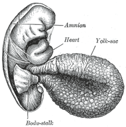
Human embryo about fifteen days old. Brain and heart represented from right side. Digestive tube and yolk sac in median section.
929:
850:
813:
695:"Toward Some Fundamentals of Fundamental Causality: Socioeconomic Status and Health in the Routine Clinic Visit for Diabetes"
299:
At the end of the fourth week, the yolk sac presents the appearance of a small pear-shaped opening (traditionally called the
524:
126:
600:
311:
as a small, somewhat oval-shaped body whose diameter varies from 1 mm to 5 mm; it is situated between the
460:, located on the opposite pole of the developing vesicle, starts its upward proliferation and meets the hypoblast.
552:
133:
219:
588:
65:
830:
498:; cells from the mesoderm pinch off an area of the yolk sac, and what remains is the secondary yolk sac.
512:
and incorporated into the embryo as the gut. The remaining part of the yolk sac is the final yolk sac.
1000:
761:
223:
187:
121:
1186:
426:
179:
109:
829:
Blaas, Harm-Gerd K; Carrera, José M (2009-01-01), Wladimiroff, Juriy W; Eik-Nes, Sturla H (eds.),
375:
Diagram showing later stage of allantoic development with commencing constriction of the yolk-sac.
1313:
953:
629:
145:
1062:
948:
564:
289:
199:
762:"Chapter One - Comparative Placental Anatomy: Divergent Structures Serving a Common Purpose"
1308:
1110:
1022:
473:
457:
230:
582:
Fetus of about eight weeks, enclosed in the amnion. Magnified a little over two diameters.
8:
1181:
965:
898:
1264:
1214:
1055:
842:
730:
722:
668:
643:
531:
880:
846:
809:
781:
714:
1126:
734:
353:
Diagram illustrating early formation of allantois and differentiation of body-stalk.
1254:
1163:
1050:
1034:
1017:
982:
970:
838:
773:
706:
663:
655:
472:: it is the vesicle which develops in the second week, its floor is represented by
210:
1323:
1229:
1210:
1176:
1082:
1012:
777:
644:"Yolk sac cell atlas reveals multiorgan functions during human early development"
495:
430:
285:
284:
and after circulating through a wide-meshed capillary plexus, is returned by the
247:
235:
114:
60:
1318:
1259:
1249:
1241:
1203:
872:
304:
293:
726:
694:
1338:
1115:
1087:
1027:
1005:
718:
546:
Human embryo—length, 2 mm. Dorsal view, with the amnion laid open. X 30.
505:
195:
659:
403:
Diagram illustrating a later stage in the development of the umbilical cord.
1224:
1072:
884:
785:
1171:
1303:
1077:
308:
274:
80:
151:
1193:
477:
453:
433:, and may be attached by a fibrous cord to the abdominal wall at the
323:. There is no clinical significance to a residual external yolk sac.
255:
251:
175:
1282:
977:
960:
943:
710:
509:
491:
320:
270:
85:
167:
1287:
1198:
316:
262:
1219:
456:
starts proliferating laterally and descending. In the meantime
312:
280:
Blood is conveyed to the wall of the yolk sac by the primitive
266:
239:
215:
171:
34:
441:
434:
281:
97:
747:
The
Developing Human: Clinically Oriented Anatomy: Chapter 7
928:
768:, Molecular Biology of Placental Development and Disease,
288:
to the tubular heart of the embryo. This constitutes the
44:
28:
766:
Progress in
Molecular Biology and Translational Science
335:
Diagram showing earliest observed stage of human ovum.
303:), into the digestive tube by a long narrow tube, the
1156:
444:
is seen opposite the site of attachment of the duct.
429:, which is situated about 60 cm proximal to the
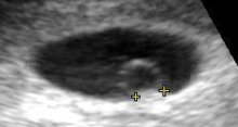
242:(measuring 3 mm as the distance between the + signs).
606:
Section through ovum imbedded in the uterine decidua
1106:
List of related male and female reproductive organs
803:
490:: this structure is formed when the extraembryonic
804:Moore, Keith; Persaud, TVN; Torchia, Mark (2013).
760:Hafez, S. (2017-01-01), Huckle, William R. (ed.),
246:The yolk sac is the first element seen within the
319:and may lie on or at a varying distance from the
273:, outside of which is a layer of extra-embryonic
1336:
504:: during the fourth week of development, during
49:Human embryo from thirty-one to thirty-four days
879:, Treasure Island (FL): StatPearls Publishing,
558:Dorsum of human embryo, 2.11 mm in length.
870:
831:"Chapter 4 - Investigation of early pregnancy"
641:
1142:
914:
828:
692:
1149:
1135:
921:
907:
642:Goh, Issac; et al. (18 August 2023).
440:Sometimes a narrowing of the lumen of the
307:. Rarely, the yolk sac can be seen in the
292:, which in humans serves as a location of
43:
27:
871:Donovan, Mary F.; Bordoni, Bruno (2020),
667:
835:Ultrasound in Obstetrics and Gynaecology
508:, part of the yolk sac is surrounded by
229:
209:
127:sac_by_E5.7.1.0.0.0.4 E5.7.1.0.0.0.4
837:, Edinburgh: Elsevier, pp. 57–78,
594:Model of human embryo 1.3 mm long.
261:The yolk sac is situated on the front (
103:vesicula umbilicalis; saccus vitellinus
1337:
930:Development of the reproductive system
693:Lutfey, Karen; Freese, Jeremy (2005).
1130:
902:
759:
755:
753:
688:
686:
516:
16:Membranous sac attached to an embryo
1345:Embryology of cardiovascular system
182:. This is alternatively called the
13:
843:10.1016/b978-0-444-51829-3.00004-0
635:
14:
1356:
1157:Membranes of the fetus and embryo
750:
683:
269:; it is lined by extra-embryonic
218:seen at approximately 5 weeks of
611:
599:
587:
575:
563:
551:
539:
523:
463:
396:
368:
346:
328:
447:
864:
822:
808:. Philadelphia, PA: Saunders.
797:
741:
234:Artificially colored, showing
214:Contents in the cavity of the
1:
699:American Journal of Sociology
677:
277:, derived from the epiblast.
778:10.1016/bs.pmbts.2016.12.001
205:
7:
1035:Definitive urogenital sinus
623:
570:Section through the embryo.
10:
1361:
530:Surface view of embryo of
480:. It is also known as the
198:during the fourth week of
1296:
1275:
1240:
1162:
1098:
1043:
1001:Development of the gonads
993:
936:
224:obstetric ultrasonography
188:Terminologia Embryologica
174:, formed by cells of the
144:
132:
120:
108:
96:
91:
79:
71:
59:
54:
42:
26:
21:
1187:Intermediate trophoblast
772:, Academic Press: 1–28,
180:bilaminar embryonic disc
660:10.1126/science.add7564
476:and its ceiling by the
873:"Embryology, Yolk Sac"
494:separates to form the
243:
227:
146:Anatomical terminology
1063:Labioscrotal swelling
496:extraembryonic coelom
427:Meckel's diverticulum
290:vitelline circulation
233:
213:
200:embryonic development
1111:Prenatal development
1023:Paramesonephric duct
806:The Developing Human
254:, usually at 3 days
1314:Reichert's membrane
1182:Syncytiotrophoblast
337:1 - Amniotic cavity
1215:Intervillous space
1056:Primordial phallus
532:Hylobates concolor
502:The final yolk sac
488:Secondary yolk sac
482:exocoelomic cavity
244:
228:
1332:
1331:
1309:Heuser's membrane
1124:
1123:
852:978-0-444-51829-3
815:978-1-4377-2002-0
517:Additional images
474:Heuser's membrane
458:Heuser's membrane
419:8 Amniotic cavity
405:1 Placental villi
385:5 Placental villi
379:2 Amniotic cavity
355:1 Amniotic cavity
301:umbilical vesicle
184:umbilical vesicle
160:
159:
155:
1352:
1255:Umbilical artery
1151:
1144:
1137:
1128:
1127:
1051:Genital tubercle
1018:Mesonephric duct
983:Cloacal membrane
971:Urogenital sinus
923:
916:
909:
900:
899:
894:
893:
892:
891:
868:
862:
861:
860:
859:
826:
820:
819:
801:
795:
794:
793:
792:
757:
748:
745:
739:
738:
705:(5): 1326–1372.
690:
673:
671:
615:
603:
591:
579:
567:
555:
543:
527:
470:Primary yolk sac
415:6 Digestive tube
409:3 Umbilical cord
400:
372:
350:
332:
166:is a membranous
152:edit on Wikidata
149:
47:
31:
19:
18:
1360:
1359:
1355:
1354:
1353:
1351:
1350:
1349:
1335:
1334:
1333:
1328:
1324:Gestational sac
1292:
1271:
1265:Wharton's jelly
1236:
1211:Chorionic villi
1177:Cytotrophoblast
1158:
1155:
1125:
1120:
1094:
1083:Vaginal process
1068:Urogenital fold
1039:
1013:Pronephric duct
989:
932:
927:
897:
889:
887:
869:
865:
857:
855:
853:
827:
823:
816:
802:
798:
790:
788:
758:
751:
746:
742:
691:
684:
680:
638:
636:Further reading
626:
619:
616:
607:
604:
595:
592:
583:
580:
571:
568:
559:
556:
547:
544:
535:
528:
519:
466:
450:
431:ileocecal valve
420:
418:
416:
414:
412:
410:
408:
406:
404:
401:
392:
390:
388:
386:
384:
382:
380:
378:
376:
373:
364:
362:
360:
358:
356:
354:
351:
342:
340:
338:
336:
333:
286:vitelline veins
248:gestational sac
238:, yolk sac and
236:gestational sac
220:gestational age
208:
170:attached to an
156:
50:
38:
17:
12:
11:
5:
1358:
1348:
1347:
1330:
1329:
1327:
1326:
1321:
1319:Vitelline duct
1316:
1311:
1306:
1300:
1298:
1294:
1293:
1291:
1290:
1285:
1279:
1277:
1273:
1272:
1270:
1269:
1268:
1267:
1262:
1260:Umbilical vein
1257:
1250:Umbilical cord
1246:
1244:
1238:
1237:
1235:
1234:
1233:
1232:
1227:
1217:
1208:
1207:
1206:
1204:Decidual cells
1196:
1191:
1190:
1189:
1184:
1179:
1168:
1166:
1160:
1159:
1154:
1153:
1146:
1139:
1131:
1122:
1121:
1119:
1118:
1113:
1108:
1102:
1100:
1096:
1095:
1093:
1092:
1091:
1090:
1085:
1075:
1070:
1065:
1060:
1059:
1058:
1047:
1045:
1041:
1040:
1038:
1037:
1032:
1031:
1030:
1025:
1020:
1010:
1009:
1008:
997:
995:
991:
990:
988:
987:
986:
985:
975:
974:
973:
968:
958:
957:
956:
951:
940:
938:
934:
933:
926:
925:
918:
911:
903:
896:
895:
863:
851:
821:
814:
796:
749:
740:
727:10.1086/428914
711:10.1086/428914
681:
679:
676:
675:
674:
637:
634:
633:
632:
630:Yolk sac tumor
625:
622:
621:
620:
617:
610:
608:
605:
598:
596:
593:
586:
584:
581:
574:
572:
569:
562:
560:
557:
550:
548:
545:
538:
536:
529:
522:
518:
515:
514:
513:
499:
485:
465:
462:
449:
446:
422:
421:
402:
395:
393:
374:
367:
365:
352:
345:
343:
334:
327:
305:vitelline duct
294:haematopoiesis
265:) part of the
207:
204:
158:
157:
148:
142:
141:
136:
130:
129:
124:
118:
117:
112:
106:
105:
100:
94:
93:
89:
88:
83:
77:
76:
73:
69:
68:
63:
61:Carnegie stage
57:
56:
52:
51:
48:
40:
39:
32:
24:
23:
15:
9:
6:
4:
3:
2:
1357:
1346:
1343:
1342:
1340:
1325:
1322:
1320:
1317:
1315:
1312:
1310:
1307:
1305:
1302:
1301:
1299:
1295:
1289:
1286:
1284:
1281:
1280:
1278:
1274:
1266:
1263:
1261:
1258:
1256:
1253:
1252:
1251:
1248:
1247:
1245:
1243:
1239:
1231:
1228:
1226:
1223:
1222:
1221:
1218:
1216:
1212:
1209:
1205:
1202:
1201:
1200:
1197:
1195:
1192:
1188:
1185:
1183:
1180:
1178:
1175:
1174:
1173:
1170:
1169:
1167:
1165:
1161:
1152:
1147:
1145:
1140:
1138:
1133:
1132:
1129:
1117:
1116:Embryogenesis
1114:
1112:
1109:
1107:
1104:
1103:
1101:
1097:
1089:
1088:Canal of Nuck
1086:
1084:
1081:
1080:
1079:
1076:
1074:
1071:
1069:
1066:
1064:
1061:
1057:
1054:
1053:
1052:
1049:
1048:
1046:
1042:
1036:
1033:
1029:
1028:Vaginal plate
1026:
1024:
1021:
1019:
1016:
1015:
1014:
1011:
1007:
1006:Gonadal ridge
1004:
1003:
1002:
999:
998:
996:
992:
984:
981:
980:
979:
976:
972:
969:
967:
964:
963:
962:
959:
955:
954:lateral plate
952:
950:
947:
946:
945:
942:
941:
939:
935:
931:
924:
919:
917:
912:
910:
905:
904:
901:
886:
882:
878:
874:
867:
854:
848:
844:
840:
836:
832:
825:
817:
811:
807:
800:
787:
783:
779:
775:
771:
767:
763:
756:
754:
744:
736:
732:
728:
724:
720:
716:
712:
708:
704:
700:
696:
689:
687:
682:
670:
665:
661:
657:
653:
649:
645:
640:
639:
631:
628:
627:
614:
609:
602:
597:
590:
585:
578:
573:
566:
561:
554:
549:
542:
537:
533:
526:
521:
520:
511:
507:
506:organogenesis
503:
500:
497:
493:
489:
486:
483:
479:
475:
471:
468:
467:
464:Modifications
461:
459:
455:
445:
443:
438:
436:
432:
428:
399:
394:
371:
366:
349:
344:
331:
326:
325:
324:
322:
318:
314:
310:
306:
302:
297:
295:
291:
287:
283:
278:
276:
272:
268:
264:
259:
257:
253:
249:
241:
237:
232:
225:
221:
217:
212:
203:
201:
197:
193:
190:(TE), though
189:
185:
181:
178:layer of the
177:
173:
169:
165:
153:
147:
143:
140:
137:
135:
131:
128:
125:
123:
119:
116:
113:
111:
107:
104:
101:
99:
95:
90:
87:
84:
82:
78:
74:
70:
67:
64:
62:
58:
53:
46:
41:
36:
30:
25:
20:
1073:Gubernaculum
949:intermediate
888:, retrieved
876:
866:
856:, retrieved
834:
824:
805:
799:
789:, retrieved
769:
765:
743:
702:
698:
651:
647:
501:
487:
481:
469:
451:
448:Histogenesis
439:
423:
383:4 Body-stalk
357:2 Body-stalk
339:2 - Yolk-sac
300:
298:
279:
260:
245:
191:
183:
163:
161:
102:
1276:Circulatory
1172:Trophoblast
534:(a gibbon).
411:4 Allantois
387:6 Allantois
359:3 Allantois
341:3 - Chorion
92:Identifiers
1304:Blastocoel
1078:Peritoneum
937:Precursors
890:2020-09-11
877:StatPearls
858:2020-10-21
791:2020-10-21
678:References
407:2 Yolk-sac
389:7 Yolk-sac
361:4 Yolk-sac
309:afterbirth
275:mesenchyme
1194:Allantois
719:0002-9602
478:hypoblast
454:hypoblast
435:umbilicus
391:8 Chorion
363:5 Chorion
256:gestation
252:pregnancy
206:In humans
176:hypoblast
81:Precursor
37:of 3.6 mm
1339:Category
1283:Placenta
1099:See also
1044:External
994:Internal
978:Ectoderm
961:Endoderm
944:Mesoderm
885:32310425
786:28110748
735:17629087
654:(6659).
624:See also
510:endoderm
492:mesoderm
417:7 Embryo
381:3 Embryo
321:placenta
315:and the
271:endoderm
192:yolk sac
164:yolk sac
86:Endoderm
22:Yolk sac
1288:Chorion
1199:Decidua
669:7614978
648:Science
413:5 Heart
377:1 Heart
317:chorion
263:ventral
250:during
186:by the
115:D015017
55:Details
1230:cavity
1220:Amnion
1164:Embryo
966:Cloaca
883:
849:
812:
784:
733:
725:
717:
666:
313:amnion
267:embryo
240:embryo
216:uterus
172:embryo
35:embryo
33:Human
1297:Other
1242:Fetus
731:S2CID
723:JSTOR
442:ileum
282:aorta
150:[
139:87180
98:Latin
881:PMID
847:ISBN
810:ISBN
782:PMID
715:ISSN
162:The
110:MeSH
72:Days
1225:sac
839:doi
774:doi
770:145
707:doi
703:110
664:PMC
656:doi
652:381
222:by
196:gut
168:sac
134:FMA
1341::
875:,
845:,
833:,
780:,
764:,
752:^
729:.
721:.
713:.
701:.
697:.
685:^
662:.
650:.
646:.
437:.
258:.
202:.
122:TE
66:5b
1213:/
1150:e
1143:t
1136:v
922:e
915:t
908:v
841::
818:.
776::
737:.
709::
672:.
658::
484:.
226:.
154:]
75:9
Text is available under the Creative Commons Attribution-ShareAlike License. Additional terms may apply.


