848:
304:
288:
276:
56:
40:
264:
333:
498:
Tears drain through the canaliculi, lacrimal sac and naso-lacrimal duct into the nose. Blockage of the naso-lacrimal duct prevents drainage of the lacrimal sac, which may lead to infection, causing a painful swelling at the side of the nose below the medial canthus. This may present as a chronically
461:
The lacrimal sac lies within a fossa in the anterior portion of the medial orbital wall. This fossa is formed by the frontal process of the maxillary bone and the lacrimal bone. The sac is surrounded by fascia, continuous with the periorbita, which runs from the anterior to the posterior lacrimal
868:
499:
watery, discharging eye, or as an acutely inflamed abscess. This should be treated with oral broad-spectrum antibiotics. The problem may recur unless drainage into the nose is re-established.
303:
129:
193:
It is oval in form and measures from 12 to 15 mm. in length; its upper end is closed and rounded; its lower is continued into the nasolacrimal duct.
379:
287:
105:
529:
491:
454:
655:
412:
803:
228:
It serves as a reservoir for overflow of tears, in which the lacrimal sac pumps inward and outward driven by the
820:
213:
522:
124:
17:
275:
888:
883:
630:
394:
342:
873:
838:
170:
733:
665:
197:
515:
220:, with surrounding connective tissue. The Lacrimal Sac also drains the eye of debris and microbes.
637:
625:
474:
Beare, Nicholas A.V.; Bastawrous, Andrew (2014). "Ophthalmology in the
Tropics and Sub-tropics".
60:
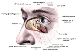
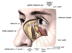
The lacrimal sac has been opened showing internal organization as well as the naso-lacrymal duct.
878:
610:
605:
425:
Anatomy: The lacrimal sac extends approximately 10 mm above the axilla of the middle turbinate.
136:
112:
100:
615:
309:
The tarsi and their ligaments. Right eye; front view. (Lacrimal sac visible at middle right.)
263:
620:
8:
743:
718:
597:
174:
847:
825:
738:
710:
483:
446:
404:
373:
320:
294:
45:
770:
723:
690:
487:
450:
408:
347:
229:
201:
178:
162:
869:
Knowledge articles incorporating text from the 20th edition of Gray's
Anatomy (1918)
760:
755:
660:
479:
442:
400:
181:, which conveys this fluid into the nasal cavity. Lacrimal sac occlusion leads to
680:
675:
670:
539:
852:
810:
728:
562:
182:
76:
862:
750:
700:
587:
582:
572:
338:
249:
245:
166:
117:
577:
557:
217:
196:
Its superficial surface is covered by a fibrous expansion derived from the
798:
142:
815:
507:
543:
467:
791:
786:
695:
685:
567:
437:
Remington, Lee Ann (2012). "Ocular Adnexa and
Lacrimal System".
386:
55:
39:
647:
71:
200:, and its deep surface is crossed by the lacrimal part of the
88:
430:
204:, which is attached to the crest on the lacrimal bone.
836:
297:. Right side. (Lacrimal sac visible at upper right.)
439:
Clinical
Anatomy and Physiology of the Visual System
363:
177:, which drain tears from the eye's surface, and the
860:
212:Like the nasolacrimal duct, the sac is lined by
473:
165:, and is lodged in a deep groove formed by the
523:
393:Sacks, Raymond; Naidoo, Yuresh (2019-01-01).
366:Sobotta's Atlas and Textbook of Human Anatomy
392:
27:Upper, dilated end of the nasolacrimal duct
530:
516:
378:: CS1 maint: location missing publisher (
54:
48:shown through dissection on the left side.
38:
436:
281:Left orbicularis oculi, seen from behind.
537:
14:
861:
357:
337:This article incorporates text in the
511:
476:Manson's Tropical Infectious Diseases
255:
656:Levator palpebrae superioris muscle
24:
484:10.1016/b978-0-7020-5101-2.00068-6
447:10.1016/b978-1-4377-1926-0.10009-8
405:10.1016/B978-0-323-47664-5.00017-1
240:The lacrimal sac can be imaged by
25:
900:
478:. Elsevier. pp. 952–994.e1.
846:
396:Endoscopic Dacryocystorhinostomy
331:
302:
286:
274:
262:
161:is the upper dilated end of the
821:Suspensory ligament of eyeball
441:. Elsevier. pp. 159–181.
214:stratified columnar epithelium
171:frontal process of the maxilla
13:
1:
364:Dr. Johannes Sobotta (1909).
326:
631:Trochlea of superior oblique
207:
188:
7:
345:of the 20th edition of
314:
223:
10:
905:
269:Medial wall of left orbit.
235:
779:
734:Accessory lacrimal glands
709:
666:Medial palpebral ligament
646:
596:
550:
248:is injected, followed by
198:medial palpebral ligament
135:
123:
111:
99:
87:
82:
70:
65:
53:
37:
32:
232:muscle during blinking.
638:Inferior oblique muscle
626:Superior oblique muscle
399:. pp. 143–148.e1.
611:Inferior rectus muscle
606:Superior rectus muscle
137:Anatomical terminology
616:Lateral rectus muscle
216:with mucus-secreting
621:Medial rectus muscle
46:lacrimal apparatusis
889:Otorhinolaryngology
884:Human head and neck
719:Lacrimal canaliculi
175:lacrimal canaliculi
711:Lacrimal apparatus
321:Lacrimal apparatus
295:lacrimal apparatus
173:. It connects the
874:Human eye anatomy
834:
833:
804:Plica semilunaris
771:Nasolacrimal duct
724:Lacrimal caruncle
691:Palpebral fissure
493:978-0-7020-5101-2
456:978-1-4377-1926-0
256:Additional images
202:orbicularis oculi
179:nasolacrimal duct
163:nasolacrimal duct
151:
150:
146:
94:saccus lacrimalis
16:(Redirected from
896:
851:
850:
842:
761:Lacrimal punctum
756:Lacrimal papilla
744:Ciaccio's glands
532:
525:
518:
509:
508:
502:
501:
471:
465:
464:
434:
428:
427:
422:
421:
390:
384:
383:
377:
369:
361:
335:
334:
306:
290:
278:
266:
242:dacrocystography
143:edit on Wikidata
140:
58:
42:
30:
29:
21:
904:
903:
899:
898:
897:
895:
894:
893:
859:
858:
857:
845:
837:
835:
830:
826:Tenon's capsule
775:
739:Krause's glands
705:
676:Meibomian gland
671:Epicanthic fold
642:
592:
546:
536:
506:
505:
494:
472:
468:
457:
435:
431:
419:
417:
415:
391:
387:
371:
370:
368:. Philadelphia.
362:
358:
332:
329:
317:
310:
307:
298:
291:
282:
279:
270:
267:
258:
238:
226:
210:
191:
147:
61:
49:
28:
23:
22:
15:
12:
11:
5:
902:
892:
891:
886:
881:
876:
871:
856:
855:
832:
831:
829:
828:
823:
818:
813:
811:Orbital septum
808:
807:
806:
796:
795:
794:
783:
781:
777:
776:
774:
773:
768:
763:
758:
753:
748:
747:
746:
741:
731:
729:Lacrimal gland
726:
721:
715:
713:
707:
706:
704:
703:
698:
693:
688:
683:
681:Ciliary glands
678:
673:
668:
663:
658:
652:
650:
644:
643:
641:
640:
635:
634:
633:
623:
618:
613:
608:
602:
600:
594:
593:
591:
590:
585:
580:
575:
570:
568:Maxillary bone
565:
563:Zygomatic bone
560:
554:
552:
548:
547:
535:
534:
527:
520:
512:
504:
503:
492:
466:
455:
429:
413:
385:
355:
354:
348:Gray's Anatomy
328:
325:
324:
323:
316:
313:
312:
311:
308:
301:
299:
292:
285:
283:
280:
273:
271:
268:
261:
257:
254:
237:
234:
225:
222:
209:
206:
190:
187:
183:dacryocystitis
149:
148:
139:
133:
132:
127:
121:
120:
115:
109:
108:
103:
97:
96:
91:
85:
84:
80:
79:
77:Angular artery
74:
68:
67:
63:
62:
59:
51:
50:
43:
35:
34:
26:
9:
6:
4:
3:
2:
901:
890:
887:
885:
882:
880:
879:Ophthalmology
877:
875:
872:
870:
867:
866:
864:
854:
849:
844:
843:
840:
827:
824:
822:
819:
817:
814:
812:
809:
805:
802:
801:
800:
797:
793:
790:
789:
788:
785:
784:
782:
778:
772:
769:
767:
764:
762:
759:
757:
754:
752:
751:Lacrimal lake
749:
745:
742:
740:
737:
736:
735:
732:
730:
727:
725:
722:
720:
717:
716:
714:
712:
708:
702:
701:Gland of Zeis
699:
697:
694:
692:
689:
687:
684:
682:
679:
677:
674:
672:
669:
667:
664:
662:
659:
657:
654:
653:
651:
649:
645:
639:
636:
632:
629:
628:
627:
624:
622:
619:
617:
614:
612:
609:
607:
604:
603:
601:
599:
595:
589:
588:Lacrimal bone
586:
584:
583:Palatine bone
581:
579:
576:
574:
573:Sphenoid bone
571:
569:
566:
564:
561:
559:
556:
555:
553:
549:
545:
541:
533:
528:
526:
521:
519:
514:
513:
510:
500:
495:
489:
485:
481:
477:
470:
463:
458:
452:
448:
444:
440:
433:
426:
416:
414:9780323476645
410:
406:
402:
398:
397:
389:
381:
375:
367:
360:
356:
353:
352:
349:
346:
344:
340:
339:public domain
322:
319:
318:
305:
300:
296:
289:
284:
277:
272:
265:
260:
259:
253:
251:
250:X-ray imaging
247:
246:radiocontrast
243:
233:
231:
221:
219:
215:
205:
203:
199:
194:
186:
184:
180:
176:
172:
168:
167:lacrimal bone
164:
160:
159:lachrymal sac
156:
144:
138:
134:
131:
128:
126:
122:
119:
116:
114:
110:
107:
104:
102:
98:
95:
92:
90:
86:
81:
78:
75:
73:
69:
64:
57:
52:
47:
41:
36:
31:
19:
18:Lachrymal sac
766:Lacrimal sac
765:
578:Ethmoid bone
558:Frontal bone
497:
475:
469:
460:
438:
432:
424:
418:. Retrieved
395:
388:
365:
359:
350:
336:
330:
241:
239:
227:
218:goblet cells
211:
195:
192:
158:
155:lacrimal sac
154:
152:
106:A15.2.07.068
93:
33:Lacrimal sac
799:Conjunctiva
244:, in which
230:orbicularis
83:Identifiers
863:Categories
816:Periorbita
420:2020-05-08
327:References
374:cite book
343:page 1028
208:Histology
189:Structure
315:See also
224:Function
853:Anatomy
792:Unibrow
787:Eyebrow
696:Canthus
686:Eyelash
598:Muscles
542:of the
462:crests.
236:Imaging
66:Details
839:Portal
661:Tarsus
648:Eyelid
490:
453:
411:
351:(1918)
72:Artery
780:Other
551:Bones
540:orbit
341:from
141:[
130:20289
89:Latin
538:The
488:ISBN
451:ISBN
409:ISBN
380:link
293:The
169:and
153:The
118:6857
101:TA98
44:The
544:eye
480:doi
443:doi
401:doi
157:or
125:FMA
113:TA2
865::
496:.
486:.
459:.
449:.
423:.
407:.
376:}}
372:{{
252:.
185:.
841::
531:e
524:t
517:v
482::
445::
403::
382:)
145:]
20:)
Text is available under the Creative Commons Attribution-ShareAlike License. Additional terms may apply.


