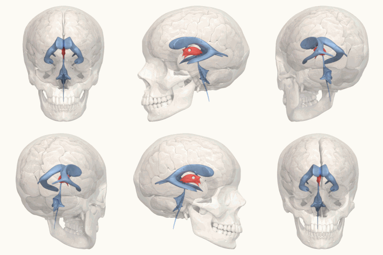
143:
131:
679:
25:
667:
691:
655:
577:. The telencephalon gradually expands laterally to a much greater extent than it does dorsally or ventrally, and its connection to the remainder of the neural tube reduces to the interventricula foramina. The diencephalon expands more evenly, but caudally of the diencephalon the canal remains narrow. The third ventricle is the space formed by the expanding canal of the diencephalon.
456:
580:
The hypothalamic region of the ventricle develops from the ventral portion of the neural tube, while the thalamic region develops from the dorsal portion; the wall of the tube thickens and becomes the hypothalamus and thalamus respectively. The hypothalamic area of the ventricle begins to distend
588:
The optic recess is noticeable by the end of the 6th week, by which time a bend is distinguishable in the dorsal portion of the ventricle border. Rostral of the bend, the medial dorsal portion of the ventrical begins to flatten, and become secretory (i.e. choroid plexus), forming the roof of the
420:
usually tunnels through the thalamic portion of the ventricle, joining together the left and right halves of the thalamus, although it is sometimes absent, or split into more than one tunnel through the ventricle; it is currently unknown whether any nerve fibres pass between the left and right
536:
form the floor posterior of the tuber cinereum, acting as the link between the fornix and the hypothalamus. Posterior of the mamillary bodies, the ventricle becomes the opening of the cerebral aqueduct, the inferior borders becoming the
569:. The lamina terminalis is the rostral termination of the neural tube. After about five weeks, different portions of the prosencephalon begin to take distinct developmental paths from one another – the more rostral portion becomes the
448:; nerve fibres reach the posterior commissure from the adjacent midbrain, but their onward connection is currently uncertain. The commissures create concavity to the shape of the posterior ventricle border, causing the
628:, particularly enlargement of the third ventricle. These observations are interpreted as indicating a loss of neural tissue in brain regions adjacent to the enlarged ventricle, leading to suggestions that
452:
above the habenular, and the deeper pineal recess between the habenular and posterior commissures; the recesses being so-named due to the pineal recess being bordered by the pineal gland.
589:
ventricle. Caudal of the bend, the ventricle border forms the epithalamus, and begins to distend towards the parietal bone (in lower vertebrates, it distends more specifically to the
408:– from the inferior side of the interventricular foramina to the anterior side of the cerebral aqueduct. The lateral border posterior/superior of the sulcus constitutes the
377:) at the posterior caudal corner. Since the interventricular foramina are on the lateral edge, the corner of the third ventricle itself forms a bulb, known as the
861:
Manji, Husseini K.; Quiroz, Jorge A.; Sporn, Jonathan; Payne, Jennifer L.; Denicoff, Kirk; Gray, Neil A.; Zarate Jr., Carlos A.; Charney, Dennis S. (April 2003).
274:
250:
1319:
1097:
486:
The portion of the floor immediately posterior of the optic chiasm distends inferiorly, and slightly anteriorly, to form a funnel (the
153:
678:
1402:
366:
863:"Enhancing neuronal plasticity and cellular resilience to develop novel, improved therapeutics for difficult-to-treat depression"
468:
89:
781:
61:
707:
862:
798:
1370:
581:
ventrally during the 5th week of development, creating the infundibulum and posterior pituitary; an outgrowth from the
68:
1204:
746:
565:. Specifically, it originates from the most rostral portion of the neural tube which initially expands to become the
108:
1090:
666:
614:
281:
42:
769:
75:
46:
269:
57:
1444:
1083:
1258:
475:
of the blood; the cerebrum lies beyond the lamina, and causes it to have a slightly concave shape. The
773:
763:
917:"Inflammation and its discontents: the role of cytokines in the pathophysiology of major depression"
815:
225:
1253:
1241:
1233:
208:
1392:
617:. An endoscopic third ventriculostomy can be performed in order to release extra fluid caused by
35:
557:
The third ventricle, like other parts of the ventricular system of the brain, develops from the
142:
810:
523:
417:
334:
257:
245:
353:
The third ventricle is a narrow, laterally flattened, vaguely rectangular region, filled with
625:
601:
The floor of the third ventricle is formed by hypothalamic structures and this can be opened
472:
401:
82:
1214:
624:
Several studies have found evidence of ventricular enlargement to be associated with major
507:
498:, which constitutes a bundle of nerve fibres from the hypothalamus. The funnel ends in the
445:
437:
1015:"Chordoid glioma: a rare radiologically, histologically, and clinically mystifying lesion"
8:
1384:
1302:
1199:
1139:
499:
491:
405:
394:
354:
327:
303:
1075:
459:
The hypothalmic portion of the third ventricle (upper right), and surrounding structures
1407:
1187:
1118:
1106:
1041:
1014:
990:
965:
941:
916:
890:
836:
712:
449:
362:
323:
307:
878:
824:
444:, which regulates sleep and reacts to light levels. Caudal of the pineal gland is the
1397:
1282:
1209:
1156:
1066:
1046:
995:
946:
882:
828:
777:
742:
633:
533:
519:
464:
370:
161:
148:
894:
1423:
1349:
1263:
1224:
1134:
1036:
1026:
985:
977:
936:
932:
928:
874:
840:
820:
690:
606:
167:
654:
1340:
1324:
1287:
1182:
640:
610:
585:(the future mouth) gradually extends towards it, to form the anterior pituitary.
506:, which is thus neurally connected to the hypothalamus via the tuber cinereum. A
503:
487:
311:
213:
177:
238:
1365:
1345:
566:
495:
390:
386:
1031:
393:; immediately above the superior central portion of the tela choroidea is the
1438:
1307:
1297:
1246:
1192:
1129:
981:
618:
570:
538:
262:
172:
130:
1177:
1050:
999:
966:"Cytokines sing the blues: inflammation and the pathogenesis of depression"
950:
886:
832:
590:
574:
480:
476:
441:
413:
315:
1149:
1110:
562:
433:
1013:
Bongetta, D; Risso, A; Morbini, P; Butti, G; Gaetani, P (28 May 2015).
426:
220:
287:
1070:
717:
582:
24:
629:
546:
409:
358:
319:
232:
684:
Drawing of a cast of the ventricular cavities, viewed from above.
602:
1067:
Stained brain slice images which include the "third%20ventricle"
432:
The posterior border of the ventricle primarily constitutes the
422:
338:
765:
First Aid for the USMLE Step 1: 2010 20th
Anniversary Edition
196:
799:"Neuroimaging studies of mood disorder effects on the brain"
514:) surrounds the superior portion of the tuber cinereum; the
479:– marks the inferior end of the lamina terminalis, with the
436:. The superior part of the posterior border constitutes the
1312:
412:, while anterior/inferior of the sulcus it constitutes the
342:
455:
1105:
1012:
361:. It is connected at the superior anterior corner to the
643:
is a rare tumour that can arise in the third ventricle.
593:); the border of the distention forms the pineal gland.
421:
thalamus via the adhesion (it has more resemblance to a
16:
Ventricle of the brain located between the two thalami
860:
739:
Textbook of
Anatomy Head, Neck, and Brain; Volume III
490:); the recess leading to the funnel is known as the
963:
914:
49:. Unsourced material may be challenged and removed.
964:Raison, C. L.; Capuron, L.; Miller, A. H. (2006).
915:Miller, A. H.; Maletic, V.; Raison, C. L. (2009).
400:The lateral side of the ventricle is marked by a
1436:
672:Coronal section of lateral and third ventricles.
636:may play a role in giving rise to the disease.
518:is in fact simply a portion of the two lateral
761:
522:, joined together by a posterior and anterior
389:, forming the inferior central portion of the
1091:
762:Le, Tao; Bhushan, Vikas; Vasan, Neil (2010).
463:The anterior wall of the ventricle forms the
741:(2nd ed.). Elsevier. pp. 386–387.
573:, while the more caudal portion becomes the
322:, in the midline between the right and left
333:Running through the third ventricle is the
1098:
1084:
141:
129:
1040:
1030:
989:
940:
814:
314:. It is a slit-like cavity formed in the
109:Learn how and when to remove this message
596:
483:forming the immediately adjacent floor.
454:
1006:
796:
385:). The roof of the ventricle comprises
1437:
180:to subarachnoid space are not visible)
1079:
736:
730:
646:
47:adding citations to reliable sources
18:
541:(sometimes historically called the
13:
1019:World Journal of Surgical Oncology
494:. The border of the funnel is the
345:that may connect the two thalami.
14:
1456:
1060:
720:line the bottom of the ventricle
689:
677:
665:
653:
615:endoscopic third ventriculostomy
282:Anatomical terms of neuroanatomy
23:
797:Sheline, Yvette (August 2003).
770:The McGraw-Hill Companies, Inc.
34:needs additional citations for
957:
933:10.1016/j.biopsych.2008.11.029
908:
854:
790:
755:
552:
440:, while more centrally it the
1:
879:10.1016/s0006-3223(03)00117-3
825:10.1016/s0006-3223(03)00347-0
724:
302:is one of the four connected
348:
171:Green – continuous with the
135:Third ventricle shown in red
7:
701:
471:monitors and regulates the
202:ventriculus tertius cerebri
10:
1461:
337:, which contains thalamic
1416:
1403:Interventricular foramina
1383:
1358:
1333:
1272:
1232:
1223:
1165:
1117:
1032:10.1186/s12957-015-0603-9
632:and related mediators of
613:in a procedure called an
381:(it is also known as the
367:interventricular foramina
280:
268:
256:
244:
231:
219:
207:
195:
190:
185:
154:interventricular foramina
140:
128:
123:
1254:Inferior medullary velum
1242:Superior medullary velum
982:10.1016/j.it.2005.11.006
158:Yellow – third ventricle
737:Singh, Vishram (2014).
460:
418:interthalamic adhesion
335:interthalamic adhesion
921:Biological Psychiatry
867:Biological Psychiatry
803:Biological Psychiatry
708:Biology of depression
597:Clinical significance
473:osmotic concentration
458:
383:bulb of the ventricle
326:, and is filled with
1215:Posterior commissure
970:Trends in Immunology
446:posterior commissure
438:habenular commissure
43:improve this article
1393:Blood–brain barrier
1385:Cerebrospinal fluid
1303:Hypoglossal trigone
1200:Hypothalamic sulcus
1183:Infundibular recess
1140:Collateral eminence
492:infundibular recess
467:, within which the
406:hypothalamic sulcus
375:aqueduct of Sylvius
355:cerebrospinal fluid
328:cerebrospinal fluid
304:cerebral ventricles
1445:Ventricular system
1408:Perilymphatic duct
1188:Suprapineal recess
1119:Lateral ventricles
1107:Ventricular system
713:Suprapineal recess
461:
450:suprapineal recess
369:, and becomes the
363:lateral ventricles
324:lateral ventricles
308:ventricular system
149:lateral ventricles
1432:
1431:
1398:Cerebral aqueduct
1379:
1378:
1283:Facial colliculus
1210:Subfornical organ
1157:Septum pellucidum
1071:BrainMaps project
783:978-0-07-163340-6
647:Additional images
634:neurodegeneration
543:cerebral peduncle
534:mammillary bodies
520:cavernous sinuses
465:lamina terminalis
371:cerebral aqueduct
296:
295:
291:
162:cerebral aqueduct
119:
118:
111:
93:
58:"Third ventricle"
1452:
1424:Ventriculomegaly
1230:
1229:
1225:Fourth ventricle
1135:Stria terminalis
1100:
1093:
1086:
1077:
1076:
1055:
1054:
1044:
1034:
1010:
1004:
1003:
993:
961:
955:
954:
944:
912:
906:
905:
903:
901:
858:
852:
851:
849:
847:
818:
794:
788:
787:
759:
753:
752:
734:
693:
681:
669:
657:
607:mamillary bodies
318:between the two
288:edit on Wikidata
285:
168:fourth ventricle
145:
133:
121:
120:
114:
107:
103:
100:
94:
92:
51:
27:
19:
1460:
1459:
1455:
1454:
1453:
1451:
1450:
1449:
1435:
1434:
1433:
1428:
1412:
1375:
1354:
1350:Lateral/Luschka
1341:Median/Magendie
1329:
1325:Sulcus limitans
1320:Medial eminence
1288:Locus coeruleus
1268:
1219:
1167:Third ventricle
1161:
1146:Occipital horn
1113:
1104:
1063:
1058:
1011:
1007:
962:
958:
913:
909:
899:
897:
859:
855:
845:
843:
816:10.1.1.597.5508
795:
791:
784:
760:
756:
749:
735:
731:
727:
704:
697:
696:Third ventricle
694:
685:
682:
673:
670:
661:
660:Third ventricle
658:
649:
641:chordoid glioma
611:pituitary gland
599:
555:
504:pituitary gland
379:anterior recess
357:, and lined by
351:
312:mammalian brain
300:third ventricle
292:
181:
175:
170:
165:
159:
157:
151:
136:
124:Third ventricle
115:
104:
98:
95:
52:
50:
40:
28:
17:
12:
11:
5:
1458:
1448:
1447:
1430:
1429:
1427:
1426:
1420:
1418:
1414:
1413:
1411:
1410:
1405:
1400:
1395:
1389:
1387:
1381:
1380:
1377:
1376:
1374:
1373:
1371:Tela choroidea
1368:
1366:Rhomboid fossa
1362:
1360:
1356:
1355:
1353:
1352:
1346:Lateral recess
1343:
1337:
1335:
1331:
1330:
1328:
1327:
1322:
1317:
1316:
1315:
1310:
1305:
1300:
1292:
1291:
1290:
1285:
1276:
1274:
1270:
1269:
1267:
1266:
1261:
1256:
1251:
1250:
1249:
1238:
1236:
1227:
1221:
1220:
1218:
1217:
1212:
1207:
1205:Tela choroidea
1202:
1197:
1196:
1195:
1190:
1185:
1180:
1171:
1169:
1163:
1162:
1160:
1159:
1154:
1153:
1152:
1144:
1143:
1142:
1137:
1132:
1123:
1121:
1115:
1114:
1103:
1102:
1095:
1088:
1080:
1074:
1073:
1062:
1061:External links
1059:
1057:
1056:
1005:
956:
927:(9): 732–741.
907:
873:(8): 707–742.
853:
809:(3): 338–352.
789:
782:
754:
747:
728:
726:
723:
722:
721:
715:
710:
703:
700:
699:
698:
695:
688:
686:
683:
676:
674:
671:
664:
662:
659:
652:
648:
645:
598:
595:
567:prosencephalon
554:
551:
525:intercavernous
516:circular sinus
512:circular sinus
500:posterior lobe
496:tuber cinereum
469:vascular organ
391:tela choroidea
387:choroid plexus
350:
347:
294:
293:
284:
278:
277:
272:
266:
265:
260:
254:
253:
248:
242:
241:
236:
229:
228:
223:
217:
216:
211:
205:
204:
199:
193:
192:
188:
187:
183:
182:
146:
138:
137:
134:
126:
125:
117:
116:
31:
29:
22:
15:
9:
6:
4:
3:
2:
1457:
1446:
1443:
1442:
1440:
1425:
1422:
1421:
1419:
1415:
1409:
1406:
1404:
1401:
1399:
1396:
1394:
1391:
1390:
1388:
1386:
1382:
1372:
1369:
1367:
1364:
1363:
1361:
1357:
1351:
1347:
1344:
1342:
1339:
1338:
1336:
1332:
1326:
1323:
1321:
1318:
1314:
1311:
1309:
1308:Area postrema
1306:
1304:
1301:
1299:
1298:Vagal trigone
1296:
1295:
1293:
1289:
1286:
1284:
1281:
1280:
1278:
1277:
1275:
1271:
1265:
1262:
1260:
1257:
1255:
1252:
1248:
1245:
1244:
1243:
1240:
1239:
1237:
1235:
1231:
1228:
1226:
1222:
1216:
1213:
1211:
1208:
1206:
1203:
1201:
1198:
1194:
1193:Pineal recess
1191:
1189:
1186:
1184:
1181:
1179:
1176:
1175:
1173:
1172:
1170:
1168:
1164:
1158:
1155:
1151:
1148:
1147:
1145:
1141:
1138:
1136:
1133:
1131:
1130:Lamina affixa
1128:
1127:
1125:
1124:
1122:
1120:
1116:
1112:
1108:
1101:
1096:
1094:
1089:
1087:
1082:
1081:
1078:
1072:
1068:
1065:
1064:
1052:
1048:
1043:
1038:
1033:
1028:
1024:
1020:
1016:
1009:
1001:
997:
992:
987:
983:
979:
975:
971:
967:
960:
952:
948:
943:
938:
934:
930:
926:
922:
918:
911:
900:September 25,
896:
892:
888:
884:
880:
876:
872:
868:
864:
857:
846:September 25,
842:
838:
834:
830:
826:
822:
817:
812:
808:
804:
800:
793:
785:
779:
775:
771:
767:
766:
758:
750:
748:9788131237274
744:
740:
733:
729:
719:
716:
714:
711:
709:
706:
705:
692:
687:
680:
675:
668:
663:
656:
651:
650:
644:
642:
637:
635:
631:
627:
622:
620:
619:hydrocephalus
616:
612:
608:
604:
594:
592:
586:
584:
578:
576:
572:
571:telencephalon
568:
564:
560:
550:
548:
544:
540:
535:
530:
528:
526:
521:
517:
513:
509:
505:
501:
497:
493:
489:
484:
482:
478:
474:
470:
466:
457:
453:
451:
447:
443:
439:
435:
430:
428:
424:
419:
415:
411:
407:
403:
398:
396:
392:
388:
384:
380:
376:
372:
368:
364:
360:
356:
346:
344:
340:
336:
331:
329:
325:
321:
317:
313:
309:
305:
301:
289:
283:
279:
276:
273:
271:
267:
264:
261:
259:
255:
252:
249:
247:
243:
240:
237:
234:
230:
227:
224:
222:
218:
215:
212:
210:
206:
203:
200:
198:
194:
189:
184:
179:
174:
173:central canal
169:
163:
155:
150:
144:
139:
132:
127:
122:
113:
110:
102:
99:November 2007
91:
88:
84:
81:
77:
74:
70:
67:
63:
60: –
59:
55:
54:Find sources:
48:
44:
38:
37:
32:This article
30:
26:
21:
20:
1178:Optic recess
1166:
1022:
1018:
1008:
976:(1): 24–31.
973:
969:
959:
924:
920:
910:
898:. Retrieved
870:
866:
856:
844:. Retrieved
806:
802:
792:
764:
757:
738:
732:
638:
623:
605:between the
600:
591:parietal eye
587:
579:
575:diencephalon
559:neural canal
558:
556:
542:
539:crus cerebri
531:
524:
515:
511:
508:venous sinus
488:infundibulum
485:
481:optic chiasm
477:optic recess
462:
442:pineal gland
431:
414:hypothalamus
399:
382:
378:
374:
352:
332:
316:diencephalon
299:
297:
251:A14.1.08.410
201:
105:
96:
86:
79:
72:
65:
53:
41:Please help
36:verification
33:
1150:Calcar avis
1111:human brain
563:neural tube
553:Development
434:epithalamus
310:within the
239:birnlex_714
191:Identifiers
725:References
626:depression
603:surgically
427:commissure
423:herniation
221:NeuroNames
69:newspapers
1334:Apertures
1264:Fastigium
1174:Recesses
811:CiteSeerX
772:pp.
718:Tanycytes
630:cytokines
583:stomodeum
545:) of the
365:, by the
349:Structure
178:apertures
166:Purple –
164:(Sylvius)
1439:Category
1247:Frenulum
1051:26018908
1000:16316783
951:19150053
895:25819902
887:12706957
833:12893109
702:See also
609:and the
547:midbrain
410:thalamus
359:ependyma
233:NeuroLex
1417:Related
1109:of the
1069:at the
1042:4453048
1025:: 188.
991:3392963
942:2680424
841:1913782
768:. USA:
561:of the
502:of the
425:than a
339:neurons
330:(CSF).
320:thalami
306:of the
214:D020542
186:Details
156:(Monro)
152:Cyan –
147:Blue –
83:scholar
1294:Lower
1279:Upper
1259:Taenia
1049:
1039:
998:
988:
949:
939:
893:
885:
839:
831:
813:
780:
745:
416:. The
404:– the
402:sulcus
395:fornix
343:fibers
160:Red –
85:
78:
71:
64:
56:
1359:Other
1273:Floor
1126:Body
891:S2CID
837:S2CID
527:sinus
510:(the
286:[
275:78454
197:Latin
90:JSTOR
76:books
1313:Obex
1234:Roof
1047:PMID
996:PMID
947:PMID
902:2013
883:PMID
848:2013
829:PMID
778:ISBN
743:ISBN
532:The
341:and
298:The
263:5769
246:TA98
209:MeSH
62:news
1348:to
1037:PMC
1027:doi
986:PMC
978:doi
937:PMC
929:doi
875:doi
821:doi
774:126
429:).
270:FMA
258:TA2
226:446
45:by
1441::
1045:.
1035:.
1023:13
1021:.
1017:.
994:.
984:.
974:27
972:.
968:.
945:.
935:.
925:65
923:.
919:.
889:.
881:.
871:53
869:.
865:.
835:.
827:.
819:.
807:54
805:.
801:.
776:.
639:A
621:.
549:.
529:.
397:.
235:ID
1099:e
1092:t
1085:v
1053:.
1029::
1002:.
980::
953:.
931::
904:.
877::
850:.
823::
786:.
751:.
373:(
290:]
176:(
112:)
106:(
101:)
97:(
87:·
80:·
73:·
66:·
39:.
Text is available under the Creative Commons Attribution-ShareAlike License. Additional terms may apply.

