264:
276:
41:
230:
at the supracondylar area of femur, where the adductor part of the adductor magnus attaches, will most likely cause damage to the femoral artery and may cause impairment of the blood supply to the lower leg. Popliteal artery can also be damaged by the fracture of distal femur.
171:
depending on whether the structure surrounding is the muscular part or the tendinous part of the adductor magnus muscle. For example, the top drawing on the right shows an oval fibrous type of adductor hiatus, and the bottom one shows a bridging muscular adductor hiatus.
29:
162:
Kale et al. classified the adductor hiatus according to its shape and the structures surrounding. An adductor hiatus is described as oval or bridging depending on the shape of the upper boundary. It can also be described as
454:
Copley, L. A.; Dormans, J. P.; Davidson, R. S. (1996). "Vascular injuries and their sequelae in pediatric supracondylar humeral fractures: toward a goal of prevention".
102:
263:
589:
524:
Wright, Lonnie B.; Matchett, W. Jean; Cruz, Carlos P.; James, Charles A.; Culp, William C.; Eidt, John F.; McCowan, Timothy C. (March 1, 2004).
45:
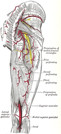
The arteries of the gluteal and posterior femoral regions. (Adductor hiatus is not labeled, but popliteal artery is visible at bottom center.)
78:
175:
Four structures are associated with the adductor hiatus. However, only two structures enter and then leave through the hiatus; namely the
1323:
275:
582:
1378:
1373:
1284:
1210:
962:
1205:
967:
249:
240:
370:
Kale, Ayşin; Gayretli, Ozcan; Oztürk, Adnan; Gürses, Ilke Ali; Dikici, Fatih; Usta, Ahmet; Sahinoğlu, Kayıhan (Dec 2012).
575:
508:
438:
311:
1253:
1024:
248:
in the relationship between the adductor hiatus and the popliteal artery can also contribute to a condition called
210:. The saphenous nerve does not leave through the adductor hiatus but penetrates superficially halfway through the
1248:
1019:
1279:
1115:
804:
33:
Deep muscles of the medial femoral region. (Adductor hiatus visible as hole in adductor magnus at lower left.)
1335:
1313:
1110:
754:
745:
97:
327:
Olson, S. A.; Holt, B. T. (Feb 1995). "Anatomy of the medial distal femur: a study of the adductor hiatus".
1039:
146:
that allows the passage of the femoral vessels from the anterior thigh to the posterior thigh and then the
301:
1006:
997:
862:
839:
678:
245:
203:
1383:
1340:
1141:
1120:
852:
782:
767:
721:
716:
1318:
1296:
1274:
1014:
794:
711:
701:
1073:
882:
777:
1410:
1301:
1215:
877:
872:
867:
827:
109:
85:
73:
498:
428:
822:
817:
772:
690:
1063:
1058:
8:
1368:
1183:
1195:
627:
623:
598:
404:
371:
196:
1149:
1029:
972:
726:
553:
545:
525:
504:
479:
471:
467:
434:
409:
391:
352:
344:
340:
307:
1159:
1154:
1127:
1085:
957:
847:
762:
567:
537:
463:
399:
383:
336:
184:
154:
and lies about 8–13.5 cm (3.1–5.3 in) superior to the adductor tubercle.
135:
1078:
933:
857:
787:
706:
671:
661:
656:
207:
164:
147:
139:
387:
1360:
923:
906:
666:
632:
211:
188:
176:
168:
151:
202:
The other two structures that are associated with the adductor hiatus are the
1404:
1105:
1068:
911:
549:
475:
395:
348:
227:
90:
269:
Schema of the arteries arising from the external iliac and femoral arteries.
918:
610:
557:
413:
180:
483:
356:
945:
541:
372:"Classification and localization of the adductor hiatus: a cadaver study"
192:
191:) immediately after they leave the hiatus, where they form a network of
115:
427:
Agur, A. M. R.; Dalley, Arthur F.; Grant, John
Charles Boileau (2013).
992:
812:
618:
602:
644:
300:
Moore, Keith L.; Dalley, Arthur F.; Agur, A. M. R. (2013-02-13).
1352:
1171:
894:
740:
143:
61:
1233:
40:
28:
369:
523:
281:
Adductor hiatus is seen as hole in the adductor magnus.
234:
453:
597:
16:
Gap between the adductor magnus muscle and the femur
526:"Popliteal Artery Disease: Diagnosis and Treatment"
496:
1402:
199:. The genicular arteries supply the knee joint.
426:
299:
497:Green, Neil E.; Swiontkowski, Marc F. (2009).
183:. Those vessels become the popliteal vessels (
583:
222:
590:
576:
39:
27:
403:
326:
217:
1403:
963:Lateral intermuscular septum of thigh
571:
433:. Lippincott Williams & Wilkins.
306:. Lippincott Williams & Wilkins.
968:Medial intermuscular septum of thigh
255:
250:popliteal artery entrapment syndrome
241:Popliteal artery entrapment syndrome
235:Popliteal artery entrapment syndrome
13:
14:
1422:
468:10.1097/00004694-199601000-00020
456:Journal of Pediatric Orthopedics
341:10.1097/00005131-199502000-00010
274:
262:
150:. It is the termination of the
517:
490:
447:
420:
363:
320:
293:
1:
329:Journal of Orthopaedic Trauma
286:
1150:Fibularis (peroneus) muscles
1030:Fibularis (peroneus) tertius
503:. Elsevier Health Sciences.
157:
7:
1324:Flexor digiti minimi brevis
500:Skeletal Trauma in Children
388:10.5152/balkanmedj.2012.030
303:Clinically Oriented Anatomy
204:descending genicular artery
10:
1427:
238:
1351:
1262:
1254:Extensor digitorum brevis
1241:
1232:
1192:
1179:
1170:
1140:
1094:
1047:
1038:
1025:Extensor digitorum longus
1005:
991:
942:
902:
893:
838:
803:
753:
739:
687:
652:
643:
609:
108:
96:
84:
72:
60:
55:
50:
38:
26:
21:
1249:Extensor hallucis brevis
1020:Extensor hallucis longus
430:Grant's Atlas of Anatomy
223:Fracture of distal femur
1280:Flexor digitorum brevis
1116:Flexor digitorum longus
1314:Flexor hallucis brevis
1285:Abductor digiti minimi
1111:Flexor hallucis longus
376:Balkan Medical Journal
126:In human anatomy, the
110:Anatomical terminology
691:Lateral rotator group
218:Clinical significance
679:Tensor fasciae latae
542:10.1148/rg.242035117
1196:Intermuscular septa
1341:Plantar interossei
1121:Tibialis posterior
853:External obturator
783:Vastus intermedius
722:External obturator
717:Internal obturator
599:Muscles of the hip
197:genicular arteries
138:(gap) between the
67:hiatus adductorius
1398:
1397:
1394:
1393:
1379:Superior extensor
1374:Inferior extensor
1336:Dorsal interossei
1319:Adductor hallucis
1297:Quadratus plantae
1275:Abductor hallucis
1228:
1227:
1224:
1223:
1136:
1135:
1015:Tibialis anterior
987:
986:
983:
982:
973:Cribriform fascia
795:Articularis genus
735:
734:
712:Superior gemellus
707:Inferior gemellus
702:Quadratus femoris
256:Additional images
124:
123:
119:
1418:
1302:Lumbrical muscle
1239:
1238:
1199:
1177:
1176:
1099:
1074:Accessory soleus
1052:
1045:
1044:
1003:
1002:
958:Iliotibial tract
949:
900:
899:
778:Vastus lateralis
751:
750:
695:
650:
649:
592:
585:
578:
569:
568:
562:
561:
521:
515:
514:
494:
488:
487:
451:
445:
444:
424:
418:
417:
407:
367:
361:
360:
324:
318:
317:
297:
278:
266:
185:popliteal artery
116:edit on Wikidata
113:
43:
31:
19:
18:
1426:
1425:
1421:
1420:
1419:
1417:
1416:
1415:
1401:
1400:
1399:
1390:
1347:
1258:
1220:
1193:
1188:
1166:
1132:
1095:
1090:
1079:Achilles tendon
1048:
1034:
996:
979:
943:
938:
934:Muscular lacuna
929:Adductor hiatus
889:
834:
828:Semimembranosus
799:
788:Vastus medialis
744:
731:
688:
683:
657:Gluteal muscles
639:
605:
596:
566:
565:
522:
518:
511:
495:
491:
452:
448:
441:
425:
421:
368:
364:
325:
321:
314:
298:
294:
289:
282:
279:
270:
267:
258:
243:
237:
225:
220:
208:saphenous nerve
160:
148:popliteal fossa
142:muscle and the
140:adductor magnus
128:adductor hiatus
120:
46:
34:
22:Adductor hiatus
17:
12:
11:
5:
1424:
1414:
1413:
1396:
1395:
1392:
1391:
1389:
1388:
1387:
1386:
1381:
1376:
1371:
1363:
1361:Plantar fascia
1357:
1355:
1349:
1348:
1346:
1345:
1344:
1343:
1338:
1328:
1327:
1326:
1321:
1316:
1306:
1305:
1304:
1299:
1289:
1288:
1287:
1282:
1277:
1266:
1264:
1260:
1259:
1257:
1256:
1251:
1245:
1243:
1236:
1230:
1229:
1226:
1225:
1222:
1221:
1219:
1218:
1213:
1208:
1202:
1200:
1190:
1189:
1187:
1186:
1180:
1174:
1168:
1167:
1165:
1164:
1163:
1162:
1157:
1146:
1144:
1138:
1137:
1134:
1133:
1131:
1130:
1125:
1124:
1123:
1118:
1113:
1102:
1100:
1092:
1091:
1089:
1088:
1083:
1082:
1081:
1076:
1071:
1066:
1055:
1053:
1042:
1036:
1035:
1033:
1032:
1027:
1022:
1017:
1011:
1009:
1000:
989:
988:
985:
984:
981:
980:
978:
977:
976:
975:
970:
965:
960:
952:
950:
940:
939:
937:
936:
931:
926:
924:Adductor canal
921:
916:
915:
914:
907:Femoral sheath
903:
897:
891:
890:
888:
887:
886:
885:
880:
875:
870:
860:
855:
850:
844:
842:
836:
835:
833:
832:
831:
830:
825:
823:Semitendinosus
820:
818:Biceps femoris
809:
807:
801:
800:
798:
797:
792:
791:
790:
785:
780:
775:
773:Rectus femoris
765:
759:
757:
748:
737:
736:
733:
732:
730:
729:
724:
719:
714:
709:
704:
698:
696:
685:
684:
682:
681:
676:
675:
674:
669:
664:
653:
647:
641:
640:
638:
637:
636:
635:
630:
615:
613:
607:
606:
595:
594:
587:
580:
572:
564:
563:
536:(2): 467–479.
516:
510:978-1416049005
509:
489:
446:
439:
419:
382:(4): 395–400.
362:
319:
312:
291:
290:
288:
285:
284:
283:
280:
273:
271:
268:
261:
257:
254:
239:Main article:
236:
233:
224:
221:
219:
216:
212:adductor canal
189:popliteal vein
177:femoral artery
159:
156:
152:adductor canal
130:also known as
122:
121:
112:
106:
105:
100:
94:
93:
88:
82:
81:
76:
70:
69:
64:
58:
57:
53:
52:
48:
47:
44:
36:
35:
32:
24:
23:
15:
9:
6:
4:
3:
2:
1423:
1412:
1411:Human anatomy
1409:
1408:
1406:
1385:
1382:
1380:
1377:
1375:
1372:
1370:
1367:
1366:
1364:
1362:
1359:
1358:
1356:
1354:
1350:
1342:
1339:
1337:
1334:
1333:
1332:
1329:
1325:
1322:
1320:
1317:
1315:
1312:
1311:
1310:
1307:
1303:
1300:
1298:
1295:
1294:
1293:
1290:
1286:
1283:
1281:
1278:
1276:
1273:
1272:
1271:
1268:
1267:
1265:
1261:
1255:
1252:
1250:
1247:
1246:
1244:
1240:
1237:
1235:
1231:
1217:
1214:
1212:
1209:
1207:
1204:
1203:
1201:
1198:
1197:
1191:
1185:
1184:Pes anserinus
1182:
1181:
1178:
1175:
1173:
1169:
1161:
1158:
1156:
1153:
1152:
1151:
1148:
1147:
1145:
1143:
1139:
1129:
1126:
1122:
1119:
1117:
1114:
1112:
1109:
1108:
1107:
1106:tarsal tunnel
1104:
1103:
1101:
1098:
1093:
1087:
1084:
1080:
1077:
1075:
1072:
1070:
1067:
1065:
1064:Gastrocnemius
1062:
1061:
1060:
1059:Triceps surae
1057:
1056:
1054:
1051:
1046:
1043:
1041:
1037:
1031:
1028:
1026:
1023:
1021:
1018:
1016:
1013:
1012:
1010:
1008:
1004:
1001:
999:
994:
990:
974:
971:
969:
966:
964:
961:
959:
956:
955:
954:
953:
951:
948:
947:
941:
935:
932:
930:
927:
925:
922:
920:
917:
913:
912:Femoral canal
910:
909:
908:
905:
904:
901:
898:
896:
892:
884:
881:
879:
876:
874:
871:
869:
866:
865:
864:
861:
859:
856:
854:
851:
849:
846:
845:
843:
841:
837:
829:
826:
824:
821:
819:
816:
815:
814:
811:
810:
808:
806:
802:
796:
793:
789:
786:
784:
781:
779:
776:
774:
771:
770:
769:
766:
764:
761:
760:
758:
756:
752:
749:
747:
742:
738:
728:
725:
723:
720:
718:
715:
713:
710:
708:
705:
703:
700:
699:
697:
694:
692:
686:
680:
677:
673:
670:
668:
665:
663:
660:
659:
658:
655:
654:
651:
648:
646:
642:
634:
631:
629:
625:
622:
621:
620:
617:
616:
614:
612:
608:
604:
600:
593:
588:
586:
581:
579:
574:
573:
570:
559:
555:
551:
547:
543:
539:
535:
531:
530:RadioGraphics
527:
520:
512:
506:
502:
501:
493:
485:
481:
477:
473:
469:
465:
462:(1): 99–103.
461:
457:
450:
442:
440:9781608317561
436:
432:
431:
423:
415:
411:
406:
401:
397:
393:
389:
385:
381:
377:
373:
366:
358:
354:
350:
346:
342:
338:
334:
330:
323:
315:
313:9781451119459
309:
305:
304:
296:
292:
277:
272:
265:
260:
259:
253:
251:
247:
242:
232:
229:
215:
213:
209:
205:
200:
198:
194:
190:
186:
182:
178:
173:
170:
166:
155:
153:
149:
145:
141:
137:
133:
132:hiatus magnus
129:
117:
111:
107:
104:
101:
99:
95:
92:
89:
87:
83:
80:
77:
75:
71:
68:
65:
63:
59:
54:
49:
42:
37:
30:
25:
20:
1330:
1308:
1291:
1269:
1194:
1096:
1049:
998:compartments
944:
928:
919:Femoral ring
746:compartments
689:
611:Iliac region
533:
529:
519:
499:
492:
459:
455:
449:
429:
422:
379:
375:
365:
335:(1): 63–65.
332:
328:
322:
302:
295:
244:
226:
201:
181:femoral vein
174:
161:
131:
127:
125:
79:A04.7.03.008
66:
1365:retinacula
1050:Superficial
946:Fascia lata
628:Psoas minor
624:Psoas major
246:Abnormality
195:called the
193:anastomoses
56:Identifiers
1216:Transverse
768:Quadriceps
727:Piriformis
287:References
1331:4th layer
1309:3rd layer
1292:2nd layer
1270:1st layer
1211:Posterior
1128:Popliteus
1086:Plantaris
1040:Posterior
848:Pectineus
813:Hamstring
805:Posterior
763:Sartorius
619:Iliopsoas
603:human leg
550:0271-5333
476:0271-6798
396:2146-3123
349:0890-5339
158:Structure
1405:Category
1369:Peroneal
1206:Anterior
1007:Anterior
863:Adductor
858:Gracilis
755:Anterior
645:Buttocks
558:15026594
414:25207041
228:Fracture
206:and the
165:muscular
1263:Plantar
1142:Lateral
883:Minimus
672:Minimus
662:Maximus
633:Iliacus
484:8747364
405:4115878
357:7714656
169:fibrous
51:Details
1384:Flexor
1353:Fascia
1242:Dorsal
1172:Fascia
1160:Brevis
1155:Longus
1069:Soleus
895:Fascia
878:Magnus
873:Brevis
868:Longus
840:Medial
667:Medius
556:
548:
507:
482:
474:
437:
412:
402:
394:
355:
347:
310:
136:hiatus
741:Thigh
144:femur
134:is a
114:[
103:58784
62:Latin
1234:Foot
1097:Deep
601:and
554:PMID
546:ISSN
505:ISBN
480:PMID
472:ISSN
435:ISBN
410:PMID
392:ISSN
353:PMID
345:ISSN
308:ISBN
187:and
179:and
91:2634
74:TA98
993:Leg
538:doi
464:doi
400:PMC
384:doi
337:doi
167:or
98:FMA
86:TA2
1407::
552:.
544:.
534:24
532:.
528:.
478:.
470:.
460:16
458:.
408:.
398:.
390:.
380:29
378:.
374:.
351:.
343:.
331:.
252:.
214:.
995:/
743:/
693::
626:/
591:e
584:t
577:v
560:.
540::
513:.
486:.
466::
443:.
416:.
386::
359:.
339::
333:9
316:.
118:]
Text is available under the Creative Commons Attribution-ShareAlike License. Additional terms may apply.

